Fewer than 10 vaquita porpoises remain in the wild, the world’s rarest marine mammal, confined to a shrinking pocket of the northern Gulf of California. Now, a team led by Florida Atlantic University has created a lasting record of the species: a high-resolution 3D digital archive of a complete vaquita skeleton, freely available to researchers, educators, and conservationists worldwide.
“This is the anatomy of a species on the edge of extinction,” says Jamie L. Knaub, a Ph.D. candidate at FAU and lead author of the study published in Marine Mammal Science. “If the worst happens, at least this data survives.”
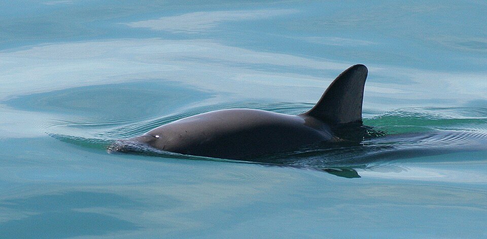
The skeleton came from a female vaquita collected in 1966, long before the species’ catastrophic decline, and donated to the San Diego Natural History Museum. The vaquita, Phocoena sinus, is the world’s smallest cetacean at about 1.5 meters (5 feet) in length. It was only described to science in 1958.
The researchers combined three imaging modalities:
- Medical CT scanning for whole-bone morphology
- Micro-CT imaging at micron resolution (finer than a human hair) for internal bone architecture
- Digital photography of each individual bone for external geometry and color
The datasets were reconstructed into interactive 3D models that can be rotated, zoomed, and measured. All data, photographs, scan datasets, and downloadable 3D meshes, have been deposited on MorphoSource, an open-access repository.
Why it matters
The vaquita’s decline has been staggering. From roughly 600 individuals in the late 1990s, the population has collapsed by over 98%, driven primarily by accidental entanglement in illegal gillnets set for totoaba, a fish whose swim bladder is trafficked to China. Mexico’s government and international conservation groups have mounted repeated rescue efforts, including a controversial “last resort” capture-and-breeding attempt in 2017 that was aborted after a captured vaquita died of stress.
With so few animals left, physical remains are irreplaceable. The original skeleton is fragile and accessible only to researchers who can visit California. The digital version removes that barrier.
“Previously, if you wanted to study vaquita anatomy, you’d need to travel to a handful of museums and handle the original bones,” says senior author Marianne E. Porter, Ph.D., also at FAU. “Now any researcher, anywhere in the world, can download millimeter-precise measurements and even 3D-print replicas for teaching.”
The open-access models also serve as a baseline for morphological comparisons with other porpoises, for evolutionary studies, and for any future conservation interventions, captive breeding or genetic rescue planning, that might require detailed anatomical knowledge.
The collaborators
The work involved the San Diego Natural History Museum (which donated the specimen), NOAA Fisheries’ Southwest Fisheries Science Center, and SeaWorld California, which co-funded the project. The full author team includes Brittany A. Dolan (FAU), Philip Unitt (San Diego Natural History Museum), Robert L. Brownell (NOAA Fisheries), and Tricia L. Meredith (FAU).
A race against extinction
“The vaquita is a symbol of what we lose when conservation fails,” says Knaub. “This digital skeleton is a record of what existed, and a tool to help the species survive.”
Whether the remaining animals can be saved remains uncertain. Gillnet bans in the vaquita’s habitat are frequently violated, enforcement is patchy, and the population may now be too small to recover. But the digital archive ensures that even if the species disappears from the wild, its biological record does not.
Source
Knaub JL, Dolan BA, Unitt P, et al. “Preserving an Imperiled Porpoise Through Pixels: Digitization of a Vaquita (Phocoena sinus) Skeleton, the World’s Most Endangered Marine Mammal.” Marine Mammal Science, 42(3) (2026). DOI: 10.1111/mms.70162
Funding: FAU’s ECOS program, Joshua M. Berlin Research Gift, FAU Laboratory Schools, SeaWorld California.