

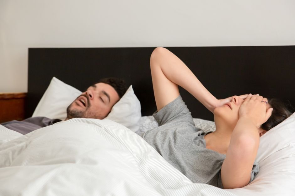
A patient walks into a prosthodontist’s office for the second time in two years. The porcelain veneer on her left molar has chipped again. The implant crown on the opposite side feels loose. She brushes twice a day, flosses, sees her general dentist every six months. By every conventional measure, her oral hygiene is impeccable. Yet her expensive restorative work keeps failing.
What her dentist may not have considered, and what a new review in the International Journal of Prosthodontics argues clinicians urgently need to start asking about, is how she sleeps at night.
The review, published online ahead of print on July 21, 2026, by researchers Francesco Buracchi, Stefano Pagano, and Nunzio Cirulli, proposes a clinical framework they call the “dual overload mechanism.” It describes how obstructive sleep apnea (OSA) may be silently accelerating the destruction of dental restorations through two converging forces: mechanical overload from nighttime teeth grinding and chemical erosion from acid reflux that occurs during apnea events.
The insight turns a familiar clinical problem on its head. For years, prosthodontists have treated failed crowns, fractured veneers, and loosening implants as isolated mechanical failures, a poorly bonded margin here, an occlusal interference there. The new review suggests many of these failures may instead carry a hidden signature of systemic disease.
The Reflex That Destroys Teeth
The mechanical half of the dual overload model centers on sleep bruxism, the rhythmic or non-rhythmic grinding and clenching of teeth during sleep. Bruxism has long been recognized as a destructive force on natural dentition and restorative materials alike. But the review repositions it within the context of OSA as a protective reflex gone awry.
When the airway collapses during an apnea event, blood oxygen drops and the brain detects a crisis. It sends an emergency signal to the jaw muscles to contract, pulling the mandible forward and the tongue away from the pharyngeal wall in a desperate attempt to reopen the airway. The resulting clench can generate forces far exceeding normal daytime mastication, forces that composite resins, ceramic veneers, and even titanium implant abutments were not designed to withstand on a nightly basis over years.
The review synthesizes evidence from the 2020-2025 literature showing that patients with moderate to severe OSA exhibit significantly higher sleep bruxism episode frequency than matched controls. More critically, the pattern of muscle activation in OSA-related bruxism differs from primary bruxism: longer sustained contractions, higher peak amplitudes, and a temporal link to respiratory event termination. Each apnea event may trigger multiple bursts of jaw clenching as the body struggles to restore airflow.
For a patient with an apnea-hypopnea index of 30 or higher, meaning their breathing stops or becomes shallow 30 or more times per hour, this cycle repeats hundreds of times a night. That is hundreds of supraphysiologic force events directed at the same vulnerable restorations, night after night, month after month.
Acid in the Dark
The chemical half of the dual overload mechanism is both more insidious and less intuitive.
Obstructive sleep apnea generates profoundly negative intrathoracic pressure during each attempted breath against a collapsed airway. Think of trying to inhale through a straw that is pinched shut. That pressure differential does not stop at the diaphragm. It extends upward, pulling gastric contents past the lower esophageal sphincter and into the pharynx and oral cavity.
Gastroesophageal reflux disease (GERD) is a well-established comorbidity of OSA, but the review reframes it as a direct pathophysiological consequence of the airway obstruction itself, not merely a coincidental finding. The chemistry of that reflux matters enormously for dental restorations. Gastric acid has a pH of approximately 1.5 to 3.5, strong enough to etch glass. When it bathes the surfaces of dental ceramics and composite resins repeatedly over months and years, it alters their surface integrity at the microscopic level.
The review cites laboratory studies showing that simulated gastric acid exposure reduces the flexural strength of lithium disilicate ceramics by 15 to 25 percent and increases surface roughness of composite resins by an order of magnitude. A surface made rougher by acid erosion provides more sites for bacterial adhesion, more friction during bruxism episodes, and more rapid propagation of subcritical cracks into outright fractures.
The dual overload, then, is not merely additive but synergistic. Acid weakens the material; clenching breaks it. Acid roughens the surface; clenching accelerates wear. Acid damages the adhesive interface between restoration and tooth; clenching peels the restoration away.
Prosthodontists as the Front Line
The review’s most provocative argument is directed at clinicians themselves. Prosthodontists, the authors contend, may be uniquely positioned to identify undiagnosed OSA earlier than sleep physicians or primary care doctors encounter these patients.
A patient presenting with recurrent restorative failures, particularly in the absence of obvious technical or hygiene-related explanations, should trigger a sleep screening. The review suggests validated tools such as the STOP-Bang questionnaire, which assesses snoring, daytime tiredness, observed apnea, blood pressure, body mass index, age, neck circumference, and male sex. A score of three or higher warrants referral for polysomnography.
The authors also flag specific intraoral signs that should raise suspicion: scalloped tongue (from nocturnal pressing against the teeth), bruxofacets on multiple restorations, unexplained marginal breakdown on crowns, and erosion patterns on the palatal surfaces of maxillary teeth, the classic “chemical striping” pattern of nocturnal reflux reaching the oral cavity.
These signs are not diagnostic of OSA on their own, but the review argues their combination should prompt a conversation that many dental clinicians are not trained to have.
Treatment Changes the Equation
There is reason for optimism, and the review documents it. Treating OSA with continuous positive airway pressure (CPAP) or mandibular advancement devices (MADs) may stabilize the oral environment in ways that extend the lifespan of dental restorations.
CPAP eliminates the negative intrathoracic pressure that drives reflux, reducing both the frequency and the acidity of nocturnal GERD episodes. Mandibular advancement devices, by repositioning the jaw forward, mechanically prevent airway collapse while simultaneously reducing the amplitude of bruxism-related muscle contractions. The review notes emerging evidence that MAD therapy, used specifically for OSA rather than as a standalone anti-bruxism device, may decrease sleep bruxism episode frequency by roughly 40 to 60 percent.
For the prosthodontist managing a patient with both OSA and recurrent restorative failures, the clinical logic becomes clear: treating the airway disease may be the single most effective intervention available for protecting the dental work.
The Bottom Line
The dual overload mechanism proposed by Buracchi, Pagano, and Cirulli reframes dental restorative failure as a potential sentinel event for undiagnosed sleep-disordered breathing. It challenges a profession rooted in mechanical solutions to think systemically, to ask about sleep quality and reflux symptoms, and to forge collaborative relationships with sleep medicine and gastroenterology colleagues.
For patients, the message is equally important. A failing crown or recurring veneer fracture is not necessarily a sign of bad dentistry. It may be a sign that something is happening in the dark, eight hours a night, that no amount of meticulous brushing can fix.
Source: Buracchi F, Pagano S, Cirulli N. Treatment of Patients with Obstructive Sleep Apnea Syndrome: What Shall Prosthodontists Consider? Int J Prosthodont. 2026 Jul 21;0(0):1-11. DOI: 10.11607/ijp.9996. PMID: 42479481.