

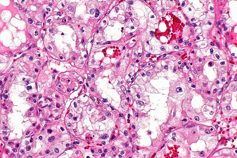
A tumor is not a uniform mass of cancer cells. It is a patchwork of regions, each with its own cellular composition, its own molecular signals, and, as a new study reveals, its own method of evading the immune system.
Researchers at the National Cancer Institute have produced the most detailed spatial map yet of HPV-associated oropharyngeal squamous cell carcinoma, a throat cancer whose incidence has been rising steadily in wealthy countries. Using a technique called Xenium spatial transcriptomics, they profiled 11 treatment-naive tumors at single-cell resolution, mapping not just which genes were active in each cell, but precisely where those cells sat within the tumor’s architecture.
The result, published in Nature Communications on June 13, reveals a tumor microenvironment organized into distinct “immune foci”, compartments that differ fundamentally in their cytokine expression, immune cell infiltration, cancer cell transcriptional states, and immunosuppressive mechanisms. In short, different regions of the same tumor fight the immune system in different ways.
The HPVoff discovery
The most surprising finding was a subpopulation of cancer cells that appear to have gone silent. These cells, which the researchers call “HPVoff”, stop expressing the viral genes that mark them as cancerous to the immune system. Because HPV-driven tumors are recognized by T cells that target viral antigens, losing HPV expression is an effective way to become invisible.
The team found these HPVoff cells scattered throughout the tumor parenchyma, but enriched in two specific environments: hypoxic (low-oxygen) zones and regions adjacent to interferon-gamma-producing T cells. Both hypoxia and IFN-γ signaling appear capable of triggering the HPVoff state, suggesting this is not a random mutation but an adaptive response: when the immune system attacks, some cancer cells simply stop displaying the target.
In laboratory assays, HPVoff cells readily evaded killing by HPV-specific T cells. “This is antigen loss as active immune evasion,” the authors suggest, a mechanism that would render any T cell-based therapy ineffective against those cells, regardless of how well checkpoint inhibitors work elsewhere in the tumor.
Why one biopsy may not be enough
The spatial compartmentalization carries a direct clinical implication. If different tumor regions rely on different resistance mechanisms, checkpoint molecule expression in one focus, immunosuppressive myeloid cells in another, antigen loss in a third, then a single biopsy could easily miss the dominant resistance strategy.
This may explain why some patients respond incompletely to checkpoint inhibitors even when their tumors are classified as immunologically “hot.” The classification may only apply to the sampled region.
The study used 10x Genomics’ Xenium platform with a custom probe set that included HPV16 genes, allowing the researchers to simultaneously measure cellular gene expression and viral transcript levels in their spatial context. The analysis was complemented by single-cell RNA sequencing of roughly 23,000 cells across 24 cell types.
Caveats
The study has significant limitations. The sample size is 11 tumors, all from a single research network within the NIH system. No independent validation cohort was analyzed. The predictive implications for checkpoint inhibitor response are inferred from the spatial patterns, not prospectively tested in patients.
The paper is also an unedited accelerated preview, the full methods, results, discussion, and figures have not yet undergone final formatting, and the journal notes that “there may be errors present.”
Several authors disclosed competing interests: Nyall R. London Jr. receives funding from Merck, and John Deeken and Gopal Bajaj receive support from Bayer, Merck, and Precision Biologics for unrelated clinical trials.
Despite these limitations, the spatial map represents a significant advance in understanding tumor heterogeneity at the level that matters most for therapy: where immune escape actually happens. The map also provides a resource for future studies, the full dataset is publicly available through GEO accession GSE290041.
Source
Sievers C, Robbins Y, Craveiro M, et al. Spatial cellular order underlies locally-confined mechanisms of immune resistance in oropharyngeal cancer. Nature Communications. 2026. DOI: 10.1038/s41467-026-74318-z