Breast compression is the most dreaded part of a mammogram. The breast is flattened between two rigid plates, sometimes with enough force to cause significant pain. Nearly one-third of women miss their initial mammogram appointment, and multiple studies have identified compression-related discomfort as a primary reason. For a screening test that reduces breast cancer mortality by 20 to 40%, that is a serious public health problem.
A new material developed at KAUST (King Abdullah University of Science and Technology) and the University of Hamburg could eliminate compression entirely, not by reducing the force applied, but by removing the need for it in the first place. The key is a copper-iodide nanocluster glass that serves as an X-ray scintillator, converting X-rays into visible light for digital detection, and is flexible enough to be moulded at just 42 degrees Celsius (107 degrees Fahrenheit), slightly above normal body temperature.
The material, described in ACS Energy Letters on May 27, 2026, achieves a spatial resolution of sub-3 micrometers (equivalent to 203 line pairs per millimeter), more than 20 times finer than current full-field digital mammography (FFDM) detectors, which typically resolve 7 to 10 line pairs per millimeter.
How it works
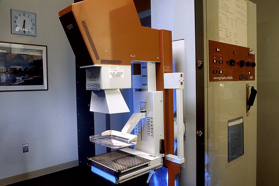
Scintillators are materials that absorb X-rays and emit visible light, which is then captured by photodetectors to form an image. In current mammography, the scintillator is a rigid flat panel, typically cesium iodide doped with thallium (CsI:Tl), that requires the breast to be flattened to uniform thickness for even exposure.
The new material is fundamentally different. It belongs to a class of zero-dimensional organic-inorganic hybrid nanocluster glasses, composed of [Cu₄I₄] cubane tetramers, four copper atoms and four iodine atoms arranged in a cube, each coordinated with organic phosphine ligands. The specific compound is [Cu₄I₄(PPh₂Et)₄]. These nanoclusters are melted and then quenched into a glass, and crucially, the individual cluster structure is preserved in the amorphous state.
The result is a free-standing glass screen with several remarkable properties: greater than 90% optical transmittance across the visible spectrum (minimizing self-absorption that degrades image quality), photoluminescence quantum yield approaching unity, and a glass transition temperature low enough, 42 degrees Celsius, that the material becomes rubbery and mouldable with gentle heating.
This last property is the one that matters clinically. Instead of a flat rigid panel, the scintillator can be formed into a curved screen that conforms to the natural shape of the breast. The breast would be imaged in its natural position, without compression. The curved detector surface eliminates the need to flatten the tissue to match a flat detector.
Resolution that reveals detail
The sub-3 micrometer resolution (203 line pairs per millimeter) is not merely an incremental improvement. Current FFDM detectors have pixel pitches of 50 to 100 micrometers (Hologic: 70 micrometers; Fujifilm: 50 micrometers); digital breast tomosynthesis systems are similar. The nanocluster glass scintillator can resolve features smaller than 3 micrometers, an improvement of roughly 20- to 30-fold in linear resolution.
In principle, this could enable detection of microcalcifications and tissue structures far smaller than what current mammography can visualize. Whether this theoretical advantage translates into earlier detection of clinically significant lesions depends on the full imaging chain, including the photodetector array that captures the scintillator’s light output, the readout electronics, and the reconstruction algorithms, none of which have been optimized for this new material.
The team demonstrated proof-of-concept by imaging test objects including a memory card, an insect, and a fish tail. These are a long way from a clinical mammogram, but the resolution metrics are unambiguous.
A decoupling that matters
Beyond the resolution and mouldability, the material embodies a fundamental discovery in scintillator physics. The researchers showed that the radioluminescence (X-ray to visible light conversion) and photoluminescence (UV to visible light conversion) pathways are kinetically and spatially decoupled in these nanocluster glasses. This means the material can achieve high scintillation efficiency independently of its photoluminescence quantum yield, breaking a long-standing assumption that good scintillators must also be good phosphors.
Practically, this decoupling means the material’s X-ray detection performance can be optimized separately from its optical properties, opening design space for even better scintillators based on the same principle.
The challenges ahead
The current work is a materials science demonstration, not a clinical device. Several major engineering challenges remain before this technology could appear in a radiology department.
First, the material must be scaled up from small test samples to full breast-sized screens while maintaining uniform thickness, optical clarity, and nanocluster integrity. Second, a completely new detector readout architecture is needed: existing flat-panel photodetector arrays cannot simply be bent to match a curved scintillator. Osman Bakr, corresponding author at KAUST, told Physics World that the next step is to “design a new detector architecture using an array of specialized optical sensors for high-resolution curved-surface imaging.”
Third, the long-term stability of the material under repeated X-ray exposure, radiation hardness, has not been characterized for clinical use. Nor have manufacturing costs, which will determine whether this approach can compete with established FFDM and tomosynthesis systems that cost $200,000 to $500,000 per unit.
One author is a founder of Quantum Solutions, a company developing quantum-dot imaging systems, representing a potential competing commercial interest.
The bigger picture
If the engineering challenges can be solved, the clinical implications extend beyond mammography. A flexible, high-resolution X-ray detector that can conform to anatomical surfaces could find applications in dental imaging, orthopedics, intraoperative imaging, and any scenario where patient comfort and image quality are both priorities.
For mammography specifically, the impact on screening compliance could be transformative. The one-third of women who skip initial mammograms represent a population at significantly elevated risk of late-stage diagnosis and death from breast cancer. A painless mammogram, the patient simply positions her breast against a warm, curved detector surface, could change screening behavior at the population level. In breast cancer screening, where every percentage point of increased compliance saves lives, that matters.
Source:
Hasanov BE, Dong C, Mohammed OF, Akturk S, Bakr OM, Bayindir M. “Nanocluster Glass Scintillators Enabling Sub-3-Micrometer Resolution and 3D Conformal X-ray Imaging.” ACS Energy Letters (2026). DOI: 10.1021/acsenergylett.6c00958