

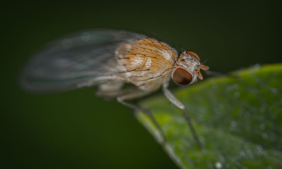
Somewhere in the genome of every fruit fly sits the fossil of an ancient virus, and a new preprint argues that this fossil is doing some of the fly’s most important night work. The Arc genes descended from retrotransposons, mobile bits of DNA that once copied themselves through animal genomes like parasites. Evolution domesticated them: the proteins they encode now assemble into hollow, virus-like shells called capsids, tiny freight containers that carry RNA between cells. A study posted to bioRxiv on August 10 reports that this ancient delivery system sets how deeply a fly sleeps, by linking two well-known groups of brain cells.
The viral inheritance
Retrotransposons are the original copy-paste machines of the genome: they copy their RNA back into DNA that inserts elsewhere, spreading relentlessly through the genomes of ancient animal ancestors. Most eventually mutated into harmless genetic clutter. Arc is the exception that got promoted. Its protein kept the ability to self-assemble into capsids, the same trick retroviruses use to build their outer shells, but traded the job of spreading infection for the job of carrying messages. Capsids package RNA, including their own messenger RNA, and transfer it into neighboring cells, a form of intercellular communication unlike the quick chemical handoffs of classical synapses.
Drosophila carries two Arc genes, dArc1 and dArc2. Earlier work tied dArc2 to long-term memory and dArc1 to how flies value sugar during learning, and in non-neuronal tissues a ligand-receptor pair called Sas and Ptp10D was shown to deliver dArc1 capsids to target cells. Whether this machinery mattered for an ongoing, hour-scale behavior remained unknown.
Deeper sleep, same rhythm
To find out, researchers used CRISPR to build flies missing dArc1, dArc2, or both genes, then tracked individuals in activity monitors for four days. Sleep in flies is defined behaviorally, as any stretch of stillness lasting five minutes or more, and by that measure the double mutants were unmistakable: across every hour of day and night, they slept far more than controls.
The extra sleep did not come from more frequent naps but from longer episodes and fewer of them, a signature of more consolidated sleep; the normal daily pattern, a daytime siesta followed by longer night sleep, stayed intact. Flies missing one Arc gene showed a milder version, mostly at night, and the double mutant’s especially long episodes suggested the two genes work together synergistically. Control experiments ruled out a movement problem: the mutants climbed and moved normally when awake. The effect was stronger in females.
Hard to wake
Sleep scientists distinguish how much an animal sleeps from how deeply. The Arc mutants are not sleeping more out of exhaustion; they are sleeping deeper. A probabilistic analysis separated the two: P(wake), the chance that a sleeping fly stirs back into activity, fell sharply in the mutants, a direct measure of depth, while sleep pressure rose only modestly at night.
The behavioral test matched the math. A low-level mechanical stimulus at the hour of peak sleep woke every control fly, but at most 59 percent of the double mutants. Mutants that did wake drifted back to sleep faster and stayed down longer. A stronger stimulus woke nearly all mutants, so sensory systems were intact; the difference was in how hard it took to rouse them. Arousal to light, however, was completely normal. Mechanical stimuli travel through the dopaminergic system, while light arousal runs through a separate cryptochrome-expressing pathway, so the selective deafness to touch pointed straight at dopamine neurons.
The neurons that matter
Two modulatory populations have long been cast as sleep’s opposing forces: serotonergic neurons promote sleep, while a group of dopaminergic cells called PAM neurons promote arousal. Knocking dArc1 down in serotonergic neurons reproduced the main mutant phenotype, longer sleep, mostly at night, with longer episodes and reduced P(wake). Knockdown in PAM neurons produced a milder version, more night sleep but no change in episode structure.
Restoring dArc1 in serotonergic neurons gave an intermediate recovery, but expressing it in PAM neurons fully restored daytime sleep, episode duration, and sleep depth to wild-type levels. Loss of the gene hit nighttime hardest; restoration worked best on daytime, a discrepancy that may reflect natural day-night variation in dArc1 expression.
The capsid requirement
The most striking control experiment targeted the capsid itself. A dArc1 variant carrying two mutations that prevent capsid assembly failed to restore sleep fully, whether delivered in serotonergic or PAM neurons. Capsid formation was needed not just to ship the message between populations but inside the receiving neurons as well. The simplest interpretation: the shell is not merely a courier, and may pass something onward from PAM neurons to downstream targets.
The delivery route: Sas to Ptp10D
How does a capsid get from a serotonin neuron to a dopamine neuron? A courier system known from non-neuronal tissues offers the route. A cell surface protein called Sas sits on the outside of extracellular vesicles, tiny membrane bubbles cells release into their surroundings. Sas binds dArc1 and steers those vesicles toward cells carrying its receptor, Ptp10D. In the fly brain, silencing dArc1 in Sas-expressing cells, or Ptp10D in PAM neurons, produced the same deeper, more consolidated nighttime sleep seen in the full mutants. The chain is coherent: serotonergic neurons pack dArc1 capsids into Sas-tagged vesicles, the vesicles dock at Ptp10D on PAM neurons, and the cargo sets how deeply the fly sleeps.
Why it matters
The finding reframes what sleep depth is. A synapse fires in milliseconds; a capsid must be assembled, loaded, shipped, and unpacked over minutes to hours. That slowness may be the point: a signal that accumulates gradually suits setting and holding a state like deep sleep, rather than triggering a quick event. There are hints the logic is ancient and general. Mice lacking Arc miss the rebound sleep that normally follows sleep deprivation and spend more time in REM sleep. Arc expression itself rises in the mouse brain after sleep deprivation, as if tracking sleep need. The conservation across species separated by hundreds of millions of years suggests the viral hand-me-down has become a core part of how brains manage sleep.
Limits
The preprint, not yet peer reviewed, leaves several threads dangling. The RNA cargo inside the capsids is unidentified. Whether endogenous dArc1 expression fluctuates across the day-night cycle remains untested. The capsid-deficient variant showed trend-level residual activity, so the requirement for capsid formation, while clear, may not be absolute. And the downstream targets of PAM neurons, and whether the capsid signals onward from them, remain open questions.
Bottom line
Sleep depth in the fruit fly appears to be set by a molecular courier system inherited from ancient viruses: capsids built by Arc proteins travel from serotonergic to dopaminergic neurons and tune how hard the sleeper is to wake. It is a reminder that the brain’s most modern-sounding functions can run on very old machinery.
Source
Butts AR, DeNiro K, Bervoets S, Shepherd JD, Caron SJC. Arc capsid signaling between serotonergic and dopaminergic neurons sets sleep depth in Drosophila. bioRxiv. 2026. doi:10.64898/2026.08.06.743357.