

Somewhere between waking and deep sleep, the brain runs a relay race it repeats every night. A slow wave of electrical silence rolls across the cortex about once a second. Close behind it, a burst of faster activity, a spindle, rises from deep inside the brain. When the two rhythms lock together in exactly the right phase, most memory researchers believe, the experiences of the day are replayed and filed into long-term storage.
That handshake has mostly been studied from the scalp, where electrodes capture when the rhythms happen but not where in the brain they matter. Now a team of researchers in Beijing has watched the whole brain at once: 107 healthy young adults, wearing caps of electrodes, falling asleep inside a functional MRI scanner. The results, published August 12 in eLife, offer the most detailed brain-wide map yet of sleep’s rhythmic machinery, and they hand the lead role to a surprising character: the thalamus, a small structure deep in the center of the brain.
A handshake in the dark
Two rhythms dominate non-REM sleep. Slow oscillations, roughly one per second, are born in the neocortex and alternate between a DOWN state, when populations of neurons fall silent, and an UP state, when they fire together. Sleep spindles, brief bursts at 11 to 16 hertz, are generated in the thalamus and ride on the UP state. Rodent experiments using optogenetics have shown that only spindles locked to the UP state in the right phase enhance memory; out-of-phase spindles do nothing. Human intracranial recordings have even suggested a triple coupling of slow oscillations, spindles, and ultra-fast hippocampal ripples, the electrical signature of memory replay. But ripples cannot be seen from the scalp, and neither EEG nor fMRI alone can tell you both when a rhythm occurs and where it acts.
107 brains on the night shift
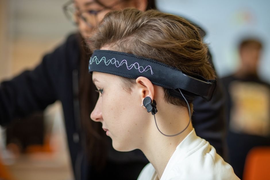
The team recruited 138 young adults (average age 22.6, 81 women) and asked them to nap in a 3-Tesla MRI scanner in the first half of the night, when deep sleep is richest. Each volunteer wore a 64-channel MR-compatible EEG cap, so the two systems recorded at once: EEG marked the exact moment of every oscillation, while fMRI photographed the blood-flow response of the entire brain at that moment. After excluding people whose heads moved too much or who slept less than ten minutes, 107 participants (63 women) remained, each contributing about three hours of sleep-scanned data. From a frontal electrode, the researchers picked out slow oscillations and spindles, then defined a coupling event as a spindle peaking within 1.5 seconds of a slow oscillation’s trough.
What the rhythms revealed
Deep sleep (stages N2 and N3) dominated the recordings, accounting for about 76 percent of sleep time, and coupled events were most common there: 2.46 per minute, versus 0.75 in light sleep and 0.43 in REM. The timing was strikingly reproducible. In 83 of the 107 participants, spindle peaks landed just before the slow oscillation’s UP-state peak, on average about 42 degrees of phase early. That offset agrees with some prior work but differs from studies that placed spindles right at the UP-state peak; detection algorithms, participant age, and subtle differences in cortical-thalamic timing could explain the gap. Coupled events also carried more punch: spindles and slow oscillations that paired up had higher amplitudes than those that went solo.
When the EEG events were aligned with the fMRI signal, each rhythm left its own footprint. Isolated slow oscillations activated the thalamus and switched off much of the neocortex, especially the default mode network, the set of regions tied to inward-directed thought. Isolated spindles lit up the thalamus, the strongest effect in the study, along with the anterior cingulate cortex and the putamen, again with the default mode network quiet. But when a spindle coupled with a slow oscillation, a new element appeared: activity in the thalamus and in the hippocampus alike, with no suppression of the default mode network. The hippocampus, seat of memory formation, woke up only during the coupled moments.
A selective relay
Using psychophysiological interaction analysis, the team asked whether communication between regions changed specifically during coupling events. Two pathways strengthened, and only then: the hippocampus talking to the thalamus, and the thalamus talking to the medial prefrontal cortex. Isolated slow oscillations and isolated spindles produced no such changes. The pattern reads like a courier service: the hippocampus sends a message, the thalamus forwards it to the cortex, and the spindle is the delivery vehicle, active only when the slow oscillation gives the green light.
As a final, open-ended probe, the team compared each rhythm’s activation pattern against NeuroSynth, a database built from thousands of published neuroimaging studies. The coupling pattern most resembled the signatures of episodic memory and declarative memory; slow oscillations leaned toward declarative memory, and spindles toward working memory. This is indirect evidence, the authors caution, because nobody in the scanner performed a memory task. Still, the resemblance is suggestive: the coupled state looks like a brain doing memory work.
Why it matters
The results reinforce a view of the thalamus as far more than a sensory relay box. During sleep it may act as the dispatcher that decides when hippocampal replay reaches the cortex. If so, spindle timing is not a curiosity; it defines the window in which memory traffic moves. The hippocampal activation during coupling also hints that ripples, invisible to scalp EEG, may be riding along, completing a triple coupling. And the disappearance of default mode suppression during coupled events suggests that the coupled state partially re-engages the networks of internally oriented cognition, consistent with memory-related reactivation.
Limits
The authors list their limits. No ripples were detected directly, so triple coupling remains an inference. All oscillations were detected from a single frontal electrode, a pragmatic choice for scanner stability that cannot capture the full spatial spread of rhythms and may underrepresent fast spindles over central and parietal areas. There was no memory task before or after sleep, so the connection to memory consolidation is inferred, not measured, and large anatomical regions of interest could hide finer effects within specific nuclei of the thalamus or subfields of the hippocampus. Follow-up work with intracranial recordings, ultra-high-field MRI, multichannel detection, and behavioral memory tests will be needed to confirm the relay model.
Bottom line
For the first time in a large human sample, the brain-wide choreography of slow oscillations and spindles has been captured in a single picture. The hippocampus proposes, the thalamus disposes, and the cortex receives, all within a second and a half. That gives sleep research a concrete target for the future, the thalamus and its spindle relay, whether the goal is closed-loop stimulation to sharpen memory or new angles on sleep-related disorders.
Source
Wang H, Zou Q, Zhang J, Gao JH, Liu Y. Human brain-wide activation of sleep rhythms. eLife. 2026;14:RP103956. doi:10.7554/eLife.103956