Asymmetric sleep spindles after thalamic stroke
brief
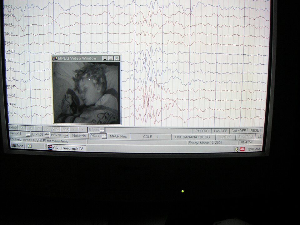
Côme-Alexandre Meyruey and Pierre Mégevand of the Neurology Division at Geneva University Hospital report a case of asymmetric sleep spindles following thalamic stroke in the June 26, 2026 online edition of *Epileptic Disorders* (DOI: 10.1002/epd2.70324). Sleep spindles, bursts of 11-16 Hz oscillatory activity characteristic of NREM sleep, are generated by thalamocortical circuits, with the thalamus serving as their primary pacemaker. A focal thalamic lesion therefore provides a natural experiment in spindle lateralization.
What it argues. The authors describe a patient in whom unilateral thalamic stroke produced a marked asymmetry in sleep spindle density and amplitude between the lesioned and intact hemispheres. Spindles were reduced or absent over the hemisphere ipsilateral to the lesion, while the contralateral side displayed preserved spindle architecture. This pattern aligns with the known anatomy of thalamocortical loop disruption: damage to the thalamic reticular nucleus or thalamocortical relay nuclei interrupts the oscillatory drive that sustains spindle generation. The case corroborates and extends earlier work from the same group on asymmetry of sleep electrophysiological markers in focal epilepsy (Sheybani et al., 2023, *Brain Communications*), where hemispheric differences in slow oscillations and spindles helped localize the epileptogenic zone.
Why it matters. Spindle symmetry is increasingly recognized as a bedside-accessible marker of thalamocortical circuit integrity. Demonstrating that a discrete thalamic infarct can produce a lateralized sleep signature has several implications. First, it refines the differential diagnosis of asymmetrical sleep EEG: a vascular thalamic lesion should now be considered alongside epilepsy as a cause of unilateral spindle loss. Second, it reinforces the value of routine sleep EEG in stroke workup; sleep architecture may reveal subclinical thalamic involvement not obvious on standard imaging or clinical examination. Third, the finding opens a translational bridge between sleep medicine and neurovascular disease: quantitative spindle metrics could serve as biomarkers for thalamic function recovery after stroke, potentially guiding rehabilitative strategies. For epilepsy clinicians already using sleep EEG for lateralization, these data provide a cautionary reminder that spindle asymmetry can also arise from an acquired structural lesion of the thalamus.
Source. Meyruey CA, Mégevand P. Asymmetric sleep spindles after thalamic stroke. *Epileptic Disord*. 2026 Jun 26. doi:10.1002/epd2.70324. PMID: 42359585. Funded by Swiss National Science Foundation grant 194507.