

Lead
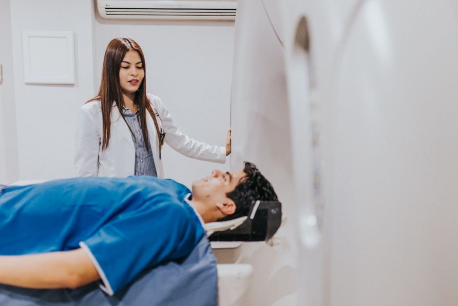
A non-invasive MRI technique that measures neuromelanin in the brainstem may help identify which patients with idiopathic REM sleep behavior disorder (iRBD) are at risk for dopamine system damage, according to a study published July 6 in Brain Imaging and Behavior. The technique could serve as a radiation-free triage tool, potentially reducing the need for costly positron emission tomography (PET) scans in patients who do not yet show signs of Parkinson disease.
Idiopathic RBD, a condition in which people physically act out their dreams during REM sleep, is one of the strongest known early markers for Parkinson disease and related synucleinopathies. Up to 80 percent of iRBD patients eventually develop a neurodegenerative condition. Dopamine transporter (DaT) imaging via PET can detect dopamine loss early, but it exposes patients to radiation and is expensive, making it impractical as a universal screening tool.
What They Found
Researchers at Samsung Medical Center and Chungnam National University in South Korea studied 66 patients diagnosed with iRBD (45 men, mean age 67) and compared them with 30 healthy controls. Using neuromelanin-sensitive MRI (NM-MRI), they measured the volume and signal properties of the substantia nigra pars compacta (SNpc), the brain region where dopamine-producing neurons reside.
Thirty-seven patients (56.1 percent) had abnormal DaT imaging results on 18F-FP-CIT PET scans and were classified as iRBD-CIT+. The key findings:
- NM-MRI metrics (volume and signal-to-noise ratio) in the SNpc were significantly reduced in iRBD-CIT+ patients compared with healthy controls, across the whole SNpc and in all three subregions (sensorimotor, associative, and limbic).
- When comparing iRBD-CIT+ directly with iRBD-CIT- patients (those with normal DaT imaging), NM volume and SNR in the associative and limbic subregions remained significantly lower in the CIT+ group, while other metrics did not differ significantly.
- Limbic subregion metrics showed the strongest discrimination between the two iRBD groups.
- Receiver operating characteristic (ROC) analysis found that NM volume had an area under the curve of 0.73, and NM SNR had an AUC of 0.75 for distinguishing iRBD-CIT+ from iRBD-CIT- patients — both in the “fair” range for a diagnostic test.
- Multivariable regression confirmed that both NM volume and SNR were independent discriminators of DaT imaging abnormality.
The study used a template-based semi-automated quantification method developed by Heuron Co., Ltd., which could allow for consistent measurements across different clinical sites.
Why It Matters
Currently, clinicians managing iRBD patients face a difficult choice: monitor patients clinically until overt Parkinson symptoms appear (by which time significant dopamine neuron loss has already occurred), or refer them for DaT imaging, which involves radiation exposure and high cost. An accessible, non-invasive biomarker could shift this paradigm.
NM-MRI adds only a few minutes to a standard MRI protocol, uses no ionizing radiation, and is already available on most clinical 3-tesla scanners. If validated in larger studies, it could function as a first-line triage tool: iRBD patients with reduced NM-MRI metrics could be prioritized for DaT PET confirmation, while those with normal NM-MRI could be spared the radiation and expense.
This matters especially as the field moves toward neuroprotective trials for Parkinson disease. Identifying iRBD patients with early dopamine dysfunction at the prodromal stage is essential for enrolling participants in clinical trials before significant neurodegeneration has occurred.
Limits
The study’s sample size is modest at 66 iRBD patients, and the findings were generated at a single academic medical center, which limits generalizability until replicated in larger, multicenter cohorts. The retrospective component of the design may introduce selection bias. Additionally, NM-MRI does not directly measure dopamine levels — it measures neuromelanin content as a proxy for the health of dopamine-producing neurons. The AUC values in the 0.73-0.75 range, while encouraging, are not yet high enough to serve as a standalone diagnostic test without follow-up confirmation.
Bottom Line
NM-MRI shows promise as a non-invasive, radiation-free method to identify which iRBD patients are most likely to have underlying dopamine transporter abnormalities. If validated, it could help clinicians triage patients for confirmatory DaT imaging and support earlier enrollment in neuroprotective clinical trials for Parkinson disease.
Source
Kim JR, Sohn B, Jo JW, Heo H, Song S, Kim EY, Joo EY. “Neuromelanin-sensitive MRI for identifying dopamine transporter imaging abnormality risk in idiopathic rapid eye movement sleep behavior disorder.” Brain Imaging and Behavior. 2026 Jul 6;20(4):106. DOI: 10.1007/s11682-026-01176-0. PMID: 42406004.