Neurotransmitter release is the fastest information-processing event in the nervous system. Within a few hundred microseconds of an action potential arriving at the synapse, synaptic vesicles fuse with the presynaptic membrane and dump their contents into the synaptic cleft. This speed is achieved by calcium sensors, proteins that detect the influx of calcium ions and trigger the fusion machinery.
For decades, the field has known that most synapses use two calcium sensors working in tandem: one for fast, synchronous release and another for slow, asynchronous release. In mammals, these are synaptotagmin-1 (Syt1) and synaptotagmin-7 (Syt7). But the precise molecular details of how they cooperate, and whether the mechanisms are the same across species, have remained unclear.
A new study published in PNAS by Lei Li, Janet Richmond, Haowen Liu, Zhitao Hu, and colleagues at City University of Hong Kong, the University of Queensland, and the University of Illinois at Chicago provides the most detailed mechanistic dissection yet of how two calcium sensors share the job. Using the nematode C. elegans, the team shows that the two sensors, SNT-1 and SNT-3, rely on overlapping but distinct molecular strategies, and that these strategies are both deeply evolutionarily conserved and subtly divergent.
Two sensors, one job
In C. elegans, the equivalent of the mammalian Syt1-Syt7 system is the SNT-1 and SNT-3 pair. SNT-1 (the fast sensor) is tethered to synaptic vesicles by an N-terminal transmembrane domain. SNT-3 (the slow sensor) is cytoplasmic, lacking this anchor. Both are synaptotagmin homologs, and together they account for essentially all evoked calcium-dependent neurotransmitter release at the C. elegans neuromuscular junction. In double mutants lacking both genes, evoked synaptic transmission is entirely abolished.
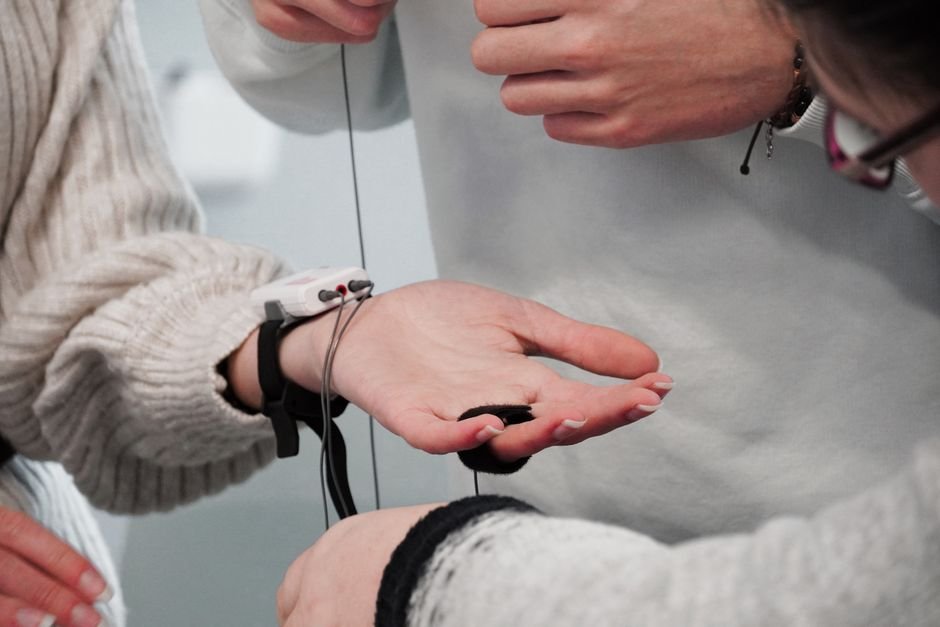
Li and colleagues used targeted mutagenesis combined with whole-cell patch-clamp electrophysiology at the nematode neuromuscular junction to systematically probe which parts of these proteins are essential. They focused on the C2 domains, calcium-binding modules that are the defining feature of the synaptotagmin family, and specifically on the interface between the C2B domain and the SNARE complex, the protein machinery that physically fuses vesicles with the plasma membrane.
The shared core
Both sensors require the same fundamental interaction. The C2B domain of each protein must bind to the SNARE complex to trigger release. Using AlphaFold 3 structural modeling, the team confirmed that the C2B-SNARE interface in SNT-1 and SNT-3 matches the canonical arrangement solved by X-ray crystallography for mammalian Syt1 (Zhou et al., Nature, 2015). This interface is evolutionarily conserved from nematodes to humans, approximately 600 million years of divergence without changing the core geometry.
Both sensors also require polybasic motifs, clusters of positively charged amino acids in their C2 domains, for membrane binding. Disrupting these motifs in either SNT-1 or SNT-3 impaired evoked release, confirming that electrostatic interaction with the plasma membrane is a conserved requirement.
Where they diverge
Despite sharing the same core interface, the two sensors use it differently.
SNT-1 engages the SNARE complex through multiple contact points, the primary interface plus additional binding sites that strengthen the interaction. This multi-pronged engagement provides the speed and reliability needed for fast synchronous release. SNT-3, by contrast, depends more restrictively on specific subregions of the C2B-SNARE interface, with less redundancy built into its binding mode.
The sensors also differ in how they handle spontaneous release, the random, calcium-independent fusion of individual vesicles that maintains baseline synaptic tone. SNT-1 mediates spontaneous release through multiple pathways, including both the primary C2B-SNARE interface and additional SNARE-binding interactions. This redundancy suggests that spontaneous release is not merely a byproduct of the evoked release machinery but a separately regulated process with its own molecular logic.
The team also discovered cell-type-specific differences. In inhibitory (GABAergic) motor neurons, secondary and tertiary regions of the C2B-SNARE interface play a more dominant role than in excitatory (cholinergic) motor neurons, compensating for the loss of polybasic-motif-mediated membrane interactions. This means the same two sensors can use different molecular strategies depending on the cell type, adding another layer of regulatory complexity.
What it means
The study establishes a framework for understanding how the brain achieves its extraordinary dynamic range of neurotransmitter release. The fast sensor (SNT-1/Syt1) provides the precise, millisecond-timed burst of release that allows the brain to compute; the slow sensor (SNT-3/Syt7) sustains release over longer timescales, contributing to short-term plasticity, the way synapses strengthen or weaken their signaling based on recent activity.
The findings also have direct relevance to neurological disease. Mutations in synaptotagmin genes are linked to epilepsy, autism spectrum disorders, intellectual disability, and movement disorders. SYT1 mutations cause a severe neurodevelopmental disorder; SYT2 mutations cause a congenital myasthenic syndrome. By mapping the precise residues and interfaces required for each sensor’s function, the study provides a molecular roadmap for interpreting disease-associated mutations, making it possible, in principle, to predict whether a particular mutation disrupts fast release, slow release, or both.
“This establishes C. elegans as a powerful model for dissecting synaptotagmin biology,” the authors write, “showing that core mechanisms are conserved from nematodes to mammals despite approximately 600 million years of divergence.”
Source:
Li L, Wang J, Xia J, Yu X, Hu J, Zhang Q, Richmond JE, Liu H, Hu Z. “Evolutionarily conserved and divergent mechanisms of dual Ca2+ sensors in synaptic vesicle exocytosis.” Proceedings of the National Academy of Sciences, Vol. 123, No. 25, e2532992123 (2026). DOI: 10.1073/pnas.2532992123