Published: June 03, 2026, 14:31 UTC
Deep brain stimulation (DBS) has been used to treat severe, treatment-resistant depression for more than two decades, with response rates of 60 to 75% in patients who have exhausted every other option. But exactly how it works has remained surprisingly unclear. DBS involves implanting electrodes in specific brain regions and delivering continuous electrical pulses. The electrodes disrupt or modulate pathological neural activity — but the clinical benefits of DBS for depression typically emerge gradually, over weeks to months, far slower than the immediate effects seen when DBS is used for Parkinson’s disease.
That slow onset has long hinted at something deeper: that DBS might not just be temporarily silencing or exciting neurons, but actually changing the brain’s physical structure.
A study published in Nature Neuroscience on June 1, 2026, provides the first direct evidence that this is exactly what happens. Researchers at the Icahn School of Medicine at Mount Sinai, led by Satoka H. Fujimoto, Atsushi Fujimoto, and co-senior authors Peter H. Rudebeck and Helen S. Mayberg, show that DBS targeting the subcallosal anterior cingulate cortex (SCC) — the standard DBS target for depression — induces white matter remodeling and remyelination in a key mood-regulating pathway.
The Target
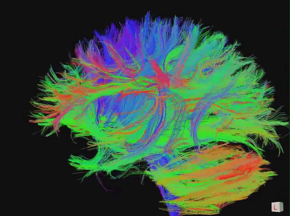
The electrode was placed at the confluence of three white matter tracts: the cingulum bundle, the forceps minor, and the uncinate fasciculus. These are the brain’s information highways connecting mood-regulating regions: the cingulum links frontal, parietal, and temporal regions; the forceps minor connects left and right prefrontal cortices; the uncinate fasciculus connects the amygdala to the orbitofrontal cortex.
For years, Helen Mayberg and her colleagues have hypothesized that DBS works by modulating activity through these pathways. But the evidence was indirect — changes in functional connectivity seen on fMRI, or metabolic changes seen on PET. The structural evidence was missing.
What They Found
The study, conducted in non-human primates (macaques) with six weeks of continuous SCC-DBS, combined in vivo neuroimaging with post-mortem histology at cellular and molecular resolution. The results were striking:
- Diffusion-weighted MRI showed a selective increase in fractional anisotropy (FA) — a measure of white matter structural integrity — specifically in the cingulum bundle, the largest of the three tracts. The other tracts were unchanged, suggesting a targeted, circuit-specific effect.
- Electron microscopy revealed the cellular basis: an increase in the number of myelinated axons and greater myelination density in the mid-cingulum bundle. The DBS was actively promoting oligodendrocyte-mediated remyelination — the formation of new myelin sheaths around axons.
- Resting-state fMRI showed that these structural changes were accompanied by brain-wide functional reorganization: altered connectivity across large-scale networks, not just locally at the stimulation site.
This is the first direct demonstration that DBS physically remodels white matter structure. The implication is profound: DBS may work, at least in part, by inducing structural plasticity — literally rewiring the brain’s communication pathways — rather than by temporarily suppressing or exciting neural activity.
Why the Slow Onset Makes Sense
If DBS works by promoting remyelination and axonal remodeling, the gradual clinical response in depression suddenly makes sense. Myelination and structural plasticity take time — days to weeks for oligodendrocyte proliferation and new myelin formation. The immediate effects seen in Parkinson’s DBS (motor improvement within seconds to minutes), by contrast, are mediated by direct modulation of neural firing rates in the basal ganglia.
This also explains why DBS for depression often requires weeks of continuous stimulation before patients report meaningful improvement, and why the benefits can persist even after stimulation is temporarily interrupted — the structural changes outlast the electrical stimulation that triggered them.
The “Pacemaker” Analogy
The study has been widely covered under the “brain pacemaker” framing — a metaphor that works on two levels. Like a cardiac pacemaker, the implantable pulse generator is placed under the collarbone with electrodes running to the target organ. And in the emerging closed-loop versions of DBS — still experimental — the device senses when the brain enters a depressive state (detected through specific neural biomarkers) and delivers stimulation only when needed, analogous to how cardiac pacemakers detect arrhythmias.
The closed-loop framing in some headlines goes beyond what the Fujimoto study actually demonstrated (the study used conventional continuous, open-loop DBS). But the conceptual shift is real: from thinking of DBS as a permanent “jamming” of pathological activity, to thinking of it as a plasticity-inducing intervention that helps the brain repair its own circuitry.
Caveats
This was a study in macaques, not humans. The SCC-DBS target was chosen because it is the established target in humans, but the stimulation parameters, duration, and electrode geometries differ. The sample size was necessarily small, as non-human primate work requires. And the study does not answer whether the white matter remodeling is causally responsible for clinical improvement — only that it is associated with DBS.
Still, for a field that has operated largely on functional imaging correlates and clinical intuition, the direct structural evidence is a major step forward. “This changes how we think about the mechanism of DBS,” Mayberg said in a statement from Mount Sinai. “It’s not just about turning a switch on and off. The brain is actively remodeling in response to the stimulation.”
Source: Fujimoto SH, Fujimoto A, Elorette C, Seltzer A, Andraka E, Holmes KR, Fleysher L, Choi KS, Russ BE, Rudebeck PH, Mayberg HS. Deep brain stimulation induces white matter remodeling and functional changes to brain-wide networks. Nature Neuroscience. 2026. DOI: 10.1038/s41593-026-02301-4. Nash Family Center for Advanced Circuit Therapeutics, Icahn School of Medicine at Mount Sinai, New York.