Published: June 03, 2026, 06:48 UTC
When patients with long COVID describe “brain fog” — the cognitive sluggishness, memory lapses, and mental fatigue that can persist for months or years after an otherwise mild infection — the leading biological hypothesis has been that the brain is simmering in a state of chronic, low-grade inflammation. Neuroinflammation seemed like a natural suspect: COVID-19 triggers a powerful inflammatory response, and lingering immune activation in the brain could explain the cognitive symptoms.
A new study published in the Journal of Neurology on April 30, 2026, puts that hypothesis to a direct test — and finds it wanting. Using the gold-standard imaging technique for detecting brain inflammation, researchers at the University of Turku in Finland found no evidence of widespread neuroinflammation in long COVID patients. The story, however, is not a simple null result. What they found instead may be more revealing.
The Gold Standard Test
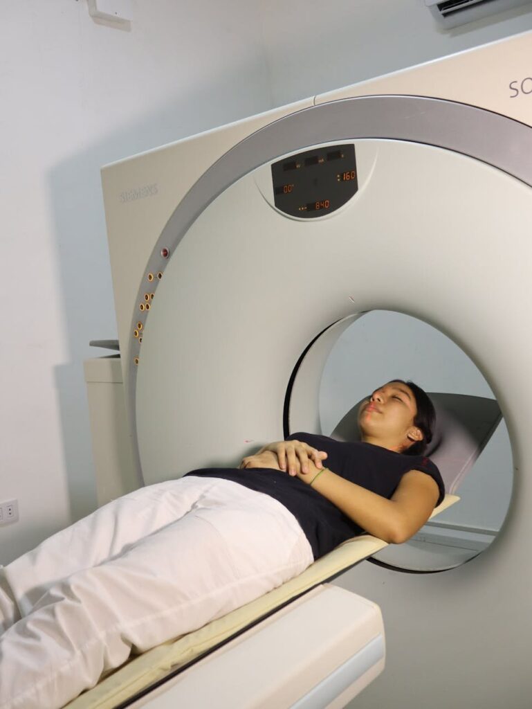
The team, led by Joel Tuomaala and Professor Laura Airas (InFLAMES Research Flagship, University of Turku), used TSPO positron emission tomography (PET) — a specialized imaging technique that detects translocator protein, a molecule expressed on activated microglia, the brain’s resident immune cells. When microglia are activated — as they are in neuroinflammatory conditions like multiple sclerosis — TSPO signal lights up on PET scans, providing a direct, quantitative measure of brain inflammation.
The study included three groups:
- 14 patients with long COVID and persistent neuropsychiatric symptoms
- 11 healthy controls with no history of COVID-19
- 13 patients with multiple sclerosis — a positive control group with known, active brain inflammation
The MS group was critical: if the TSPO-PET method failed to detect inflammation in known MS lesions, any negative result in the long COVID group would be uninterpretable.
The Result: No Widespread Inflammation
The primary finding was unambiguous: “We did not observe evidence of widespread brain inflammation in patients with long COVID when compared to healthy controls.” The long COVID group showed no significant increase in TSPO signal across the brain as a whole. The MS group, by contrast, showed the expected elevated TSPO signal — confirming the imaging method worked correctly and ruling out a false negative.
This result directly contradicts the widespread neuroinflammation hypothesis that has dominated long COVID research for years. If brain fog were driven by diffuse, ongoing inflammation, it should have been detectable with TSPO-PET, the most sensitive clinical tool available. It was not.
What They Found Instead
But the analysis did not stop at the global level. When the researchers examined specific brain regions, a different picture emerged. They found increased TSPO signal — indicating persistent glial activation — localized to the limbic system, specifically the thalamus, amygdala, and hippocampus.
These are not random structures. The limbic system is the brain’s emotional and memory network. The amygdala processes fear and threat. The hippocampus is central to memory formation. The thalamus relays sensory and motor signals. Patients with the most severe fatigue, anxiety, and depression showed the strongest limbic TSPO signal — a dose-response relationship that suggests the glial activation is clinically relevant.
The pattern was also temporally dynamic. Patients scanned within 16 months of their initial infection showed higher inflammatory activity than those whose symptoms had persisted longer, suggesting that any COVID-related neuroglial activation may resolve over time rather than persist indefinitely.
What This Means
The study redirects the long COVID research agenda in two important ways.
First, it rules out the simplest model — that long COVID brain fog is just “COVID inflammation in the brain.” That hypothesis can now be set aside, and resources redirected toward alternative mechanisms: vascular damage, autonomic dysfunction, metabolic changes in neurons, or the limbic-specific glial activation the Turku team actually observed.
Second, the limbic system finding is itself a new hypothesis. If glial activation is concentrated in emotion- and memory-processing circuits, it may explain why long COVID cognitive symptoms are so intertwined with mood disorders — anxiety, depression, and emotional dysregulation are among the most disabling features for many patients. The brain fog may not be a diffuse cognitive deficit but a disruption of the circuits that regulate attention, emotional context, and memory consolidation.
Limitations
The sample size is small — 14 long COVID patients — and the findings need replication in larger, more diverse populations. TSPO-PET is expensive and requires specialized radiochemistry, which limits its widespread use. The study cannot distinguish whether the limbic glial activation is a cause of symptoms or an epiphenomenon of some other underlying process. And the study cannot rule out the possibility that neuroinflammation occurs at a level below PET detection, or that it occurs in cellular populations (such as astrocytes) that TSPO does not capture well.
Still, for the tens of millions of people worldwide living with long COVID cognitive symptoms, the study provides something the field has lacked: a clear, testable, negative result that narrows the search space — and a specific new target for investigation.
Source: Tuomaala J, Saraste M, Smith E, Kuusi M, Westerberg E, Honkonen E, Kargar R, Laaksonen S, Lehto J, Luoma A, Matilainen M, Misin O, Atosuo J, Airas L, et al. Association between post-COVID-19 neuropsychiatric symptoms and persistent glial activation in the limbic system: a TSPO PET study. Journal of Neurology. 2026. DOI: 10.1007/s00415-026-13842-w. University of Turku & Turku PET Centre, Turku University Hospital, Finland.