

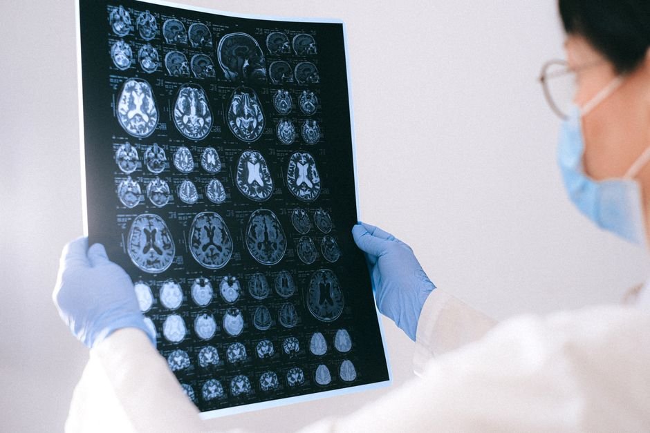
For three decades, researchers have tried to see what the brain does during sleep, not just which electrical rhythms pulse across the scalp, but which deep structures light up and fall silent across the night. The challenge has been technical: electroencephalography (EEG) captures millisecond-scale changes in cortical activity but cannot localize them subcortically, while functional neuroimaging methods such as fMRI and PET offer spatial precision at the cost of temporal resolution. Combining them simultaneously has promised the best of both worlds, but the literature has grown in scattered directions, and no one had assembled the full picture until now.
The first systematic review to span the entire field of simultaneous EEG-neuroimaging sleep research has been published in Neuroimage. Led by Angeliki Pollatou of the Uniformed Services University and Walter Reed National Military Medical Center, the review draws on systematic searches across five databases and identifies 205 eligible studies published over roughly 30 years. The result is a comprehensive map of convergent brain activity patterns during sleep, paired with a sharp-eyed accounting of the methodological heterogeneity that still limits the field.
Across modalities, a convergent picture emerges
The included studies break down by imaging modality: EEG-fMRI dominates with 141 studies, followed by EEG-PET (34 studies), EEG-NIRS (26 studies), and multimodal combinations (4 studies). The diversity of methods is both a strength and a challenge, but the authors find striking convergence where the data is most robust.
For non-REM (NREM) sleep, the evidence converges across modalities on the involvement of three key regions: the thalamus, the precuneus, and the cingulate cortex. These structures form a core network whose coordinated activity characterizes the NREM state, supporting the longstanding view of NREM sleep as a period of thalamocortical synchronization and reduced sensory throughput.
For REM sleep, the pattern shifts dramatically. Limbic and paralimbic networks, including the amygdala and hippocampus, show increased activity, consistent with the emotional and memory-processing functions long attributed to REM. At the same time, executive control regions, particularly the dorsolateral prefrontal cortex, show reduced activity. This dissociation between emotional brain regions and prefrontal regulatory circuits may help explain the vivid, emotionally charged, and often illogical quality of dreaming.
Methodological heterogeneity: a field still finding its footing
The review’s most candid contribution may be its systematic portrait of how heterogeneous the field remains. Among the 205 studies, 49 percent were overnight-only protocols, 20 percent used nap-only designs, and 74 percent enrolled only healthy adults. The median sample size was just 18 participants. Sixty-one percent of studies used the standard 30-second epoch for sleep staging, but the remainder adopted varied approaches, creating friction for cross-study comparison. Twenty-eight percent used sleep restriction protocols before imaging, further complicating generalization.
This heterogeneity matters because it shapes what can and cannot be concluded. Small samples limit statistical power and raise the risk of false positives. Varied staging approaches mean that what counts as NREM or REM may differ subtly from study to study. And the heavy reliance on healthy adults leaves open the question of how these circuits behave in clinical populations where sleep is disrupted: insomnia, sleep apnea, PTSD, and neurodegenerative disorders.
A reference for the next generation of sleep neuroimaging
The review’s primary value is as a landmark synthesis. By bringing three decades of work under one roof and identifying where findings converge and where they diverge, Pollatou and colleagues have created a reference framework that future studies can build on. The convergent findings, thalamus and cingulate in NREM, limbic activation and prefrontal deactivation in REM, represent the field’s most robust discoveries, validated across multiple imaging modalities and independent laboratories.
The authors also flag the need for larger samples, standardized protocols, and more diverse populations. As simultaneous EEG-neuroimaging becomes more accessible through advances in MRI-compatible EEG systems and portable NIRS devices, the field has an opportunity to move from small, single-site studies to larger, multi-site collaborations that can deliver the statistical power and generalizability that sleep neuroscience requires.
The conflicts of interest statement notes that Nastaren Abad, Afis Ajala, and Luca Marinelli are employees of GE HealthCare, which has commercial interests in neuroimaging technology. All other authors declare no competing interests.
Source
Pollatou A, Tays G, Skeiky L, Coon W, Scholl C, Abad N, Metzger E, Marinelli L, Ajala A, Yeh PH, Doty TJ, Shih R, Lee A, Capaldi VF II, Kim S, Werner JK. Mapping Human Sleep-State Brain Function with Simultaneous EEG and Neuroimaging: A systematic review. Neuroimage. 2026 Jul 10:122122. doi: 10.1016/j.neuroimage.2026.122122. PMID: 42431562.